1. PICASSO LASER MACHINE
Laser (Light Amplification by Stimulated Emission of Radiation) was invented in 1960. Up to now, after more than 50 years of development, laser has been widely applied in many fields of science, technology and life. In the medical industry in general and dentistry in particular, although only recently developed, the great influence of laser is revolution in diagnosis and treatment thanks to its superior features. One of the new trends in dentistry is the application of Laser in dental treatment. Using laser in dental treatment supports dentists, reduces stress and pain for patients; in addition, it also supports dentists in performing minimally invasive dental interventions, especially for children in hard tissue and soft tissue surgery with little discomfort, no pain during and after treatment, no infection and little or no bleeding.
Application of laser in soft tissue treatment
Tongue-tie, low labial frenulum, tooth exposure for orthodontic treatment; gingival contouring surgery and removal of damaged gingival tissue for orthodontic patients, crown lengthening in the treatment of tooth decay; biopsy; treatment of complications due to tooth eruption; treatment of Aphtes and Herpes lesions.
- Application of laser in pulp treatment: For vital teeth, the laser is set at 20-30hz to clean the pulp cavity in 10-20s.
- Application in tooth whitening: Soft tissue low-intensity laser can be used to speed up the tooth whitening process.
- Application in cutting benign tumors
2. EQ-V THERMAL MACHINE
Root canal obturation is one of the key factors for successful endodontic treatment (root canal treatment).
EQ-V is a wireless thermal obturation device, applying continuous wave (CW- Continuous Wave) and stable heat transfer technology, helping to avoid sudden temperature changes affecting the tissue around the apex.
With a new technology pump head, this device helps to deliver root canal filling material (Gutta Percha) to most of the lateral canals and their extensions, while also ensuring the sealing of the apical 1/3 and accurate.
This device uses the vertical method, which is capable of filling the root canal in 3 dimensions, which is a condition to help achieve long-term root canal treatment results.
3. PULP TESTER
Sakura Dental uses the Gentle Pulp Tester to aid in diagnosis by applying a stimulus in the form of an electrical pulse to the tooth being tested to evaluate the responsiveness of the sensory nerves inside the root canal. This way, it is possible to determine whether the pulp is alive or not.
This test is very important in diagnosis, because It helps to accurately determine the level of inflammation, partial or complete death of the dental pulp, avoiding root canal treatment if not really necessary.
4. CT SCAN
Sakura Dental Clinic applies modern machinery in diagnosis and treatment, including the use of CT Scan. Cone beam CT creates 3D images of tooth structure, tissue soft tissue, nerves, and bones of the maxillofacial region allowing for precise treatment planning. Compared with traditional dental X-rays, this method Cone beam CT X-ray method brings many outstanding advantages such as:
- The X-ray beam is more focused, so it gives better quality 3D images than traditional dental X-ray methods.
- Provides many different angles with just one scan.
- This method is painless, non-invasive and for absolutely accurate results.
NewTom GO 3D CT application at Sakura Dental Clinic:
- Detecting pathologies in the jawbone: CBCT helps detect, diagnose, and assess the spread of invasive jawbone pathologies such as ameloblastoma, odontogenic cyst, jawbone cancer, etc., thereby helping to screen and timely advice to customers.
- Detect bone fractures and tooth fractures due to trauma: Tooth trauma is a common type of trauma in accidents involving the head and face area that 2D films may not clearly show the path and direction of the fracture. Similar to bone fractures, CBCT films will most clearly show the fracture paths and bone fragments so that the doctor can plan appropriately and most aesthetic.
- Application in orthodontics: With CBCT films, orthodontists can see the status of impacted teeth, the direction of tooth growth, the correlation and development of the jaw bone, the position of the bone to place miniscrews, analyze orthodontic parameters on facial aesthetics, evaluate the results before and after orthodontic treatment of the patient.
- CBCT in Implant treatment planning uses 3D images and multi-slice to accurately determine the height, width and anatomy of the jawbone and alveolar bone as well as the relationship of the edentulous area to adjacent anatomical structures such as the inferior alveolar canal.
Implant placement with surgical guides can also be performed with CBCT data.
Thanks to the 3D capabilities of CBCT, our doctors can decide whether need bone grafting, sinus lift before implant placement or not, as well as choosing the most suitable implant size for each bone area.
- CBCT in diagnosing impacted/ectopic wisdom teeth: Thanks to CBCT, Dr. Sakura has can predict the relationship of the nerve to the wisdom tooth root as well as decide to perform the wisdom tooth extraction technique in one time or just cut the wisdom tooth body, waiting for the tooth root to emerge.
- CBCT in endodontic treatment: In root canal treatment or endodontic treatment, complex anatomical structures of the root canal such as deep division of the root canal, branching root canal, accessory root canal, C-shaped root canal and pathological conditions of the root canal such as root stones, root canal calcification cause great difficulty in treatment and cannot be seen on conventional 2D X-ray films.
With CBCT films, it will provide maximum support for doctors in evaluating the anatomy of the root canal in 3D space as well as analyzing obstacles and root canal pathologies, thereby supporting the treatment process to be effective and safe.
- CBCT in diagnosing temporomandibular joint pathology: CBCT films visually capture the temporomandibular joint (TMJ) on each side with can be used to assess changes in the bony part of this region to the mandibular condyle and temporal bone as well as the relationship of the ball and socket in the joint. Changes in this bony part can be the result of trauma/bone damage, dysplasia, hyperplasia or bone pathology
5. ITERO SCAN MACHINE
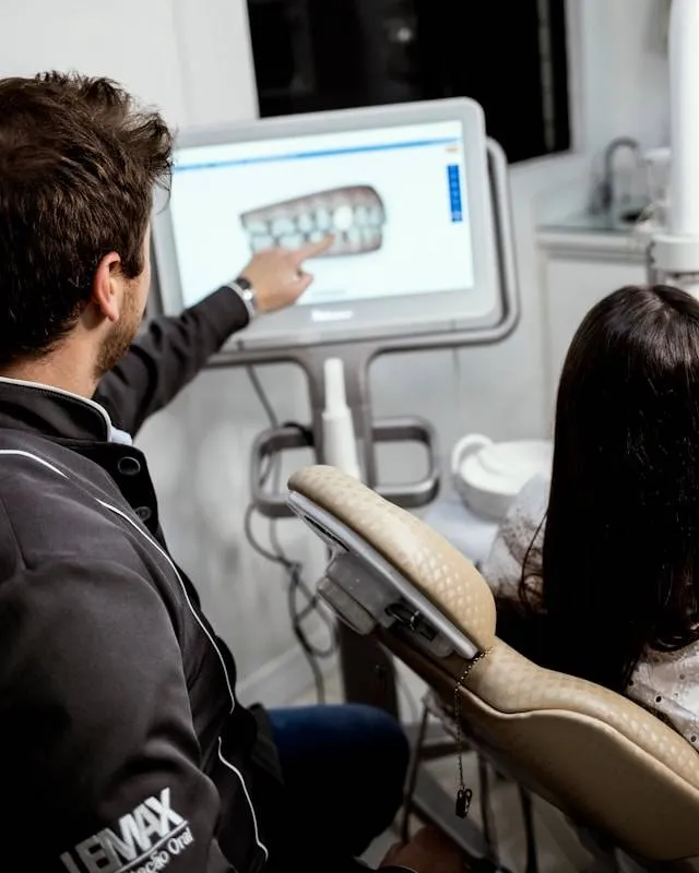
Itero isve; dental impression scanning device with integrated AI artificial intelligence technology to provide doctors with the most detailed and realistic images of teeth and gums, even in the most hidden and difficult to observe positions. This technology is considered the pinnacle because it helps to completely overcome the disadvantages of the classic impression technique.
ITERO laser jaw scanner produces multidimensional images of teeth, oral tissue, jaw structure, and bite. Thanks to that, the doctor knows exactly the oral health status of the patient and make decisions and treatment plans.
In addition, the parameters of the iTero 5D dental impression machine after scanning are stored in the iTero machine and can be transmitted to the computer to design orthodontic devices and precise restorations. The data is also transmitted to the ClinCheck software to simulate the treatment process and future dental results, after the process. orthodontic treatment.
5.1. Niri infrared technology
Niri is a modern technology that is far superior to old technologies. Niri allows:
- See images inside the tooth.
- Visualize the structure inside the tooth in the most intuitive way.
- Doctors can easily diagnose the patient's condition.
- Help doctors plan treatment quickly and most accurate.
- Can detect early dental diseases before we can see them on X-rays.
5.2 Evaluation orthodontic progress through iTero Element 5D
In addition to scanning teeth impressions, iTero 5D is superior because it allows doctors to evaluate the patient's progress. This feature is very interesting, it can accurately compare the Invisalign treatment stage with the previous simulation in ClinCheck software. This is very useful for doctors to continue to have adjustments in treatment planning if necessary.
5.3 Invisalign Outcome Simulator Description
ITero Element 5D Outcome Simulator is a feature that uses scans to allow patients to see the process of tooth movement while wearing Invisalign trays and the final result after orthodontic treatment. Thanks to that, customers can be completely assured because there is can track and monitor the progress of teeth moving little by little from the first aligner tray until the end of the teeth adjustment process.
5.4 TimeLapse feature helps predict problems
When learning about iTero 5D, we will know about the TimeLapse feature of the iTero Element 5D dental impression scanner based on the results of each scan can predict problems. This feature helps:
- Better see and understand how soft tissue is receding.
- Movement or any wear or tear of the tooth.
- Can see changes whether minimum after each treatment.
6. PIEZOTOME BONE SURGERY MACHINE
It is time for atraumatic implant surgery. 50% of all implants placed require graftingbone e;p. Perform implant surgery at the same speed as rotary instruments with greater safety & less trauma.


